• Skip to primary navigation
  • Skip to main content
  • Skip to primary sidebar
  • Skip to footer

Johns Hopkins Medicine Lyme Disease Research Center

Show Search
Hide Search
  • Lyme Disease
    • Signs & Symptoms of Lyme Disease
    • Diagnosis of Lyme Disease
    • Lyme Disease Treatment and Prognosis
    • Lyme Disease Prevention, Tick Removal, & Health and Wellness Tips
    • Frequently Asked Questions (FAQs)
    • Lyme Disease Video Introduction Series
    • Lyme Disease Video Education Resources
  • About the Center
    • Meet Our Team
    • Make an Appointment
    • Address & Directions
    • Refill a Prescription
    • Lyme and Tickborne Disease Fellowship Program
    • Subscribe to Our Email Newsletter
  • News & Impact
    • Impact Report
    • Newsletters
    • Research News
    • In The News
    • Talks by John Aucott, MD
    • Subscribe to Our Email Newsletter
  • Research Studies
    • Open Research Studies
    • Clinical Trials Network for Lyme and Other Tick-Borne Diseases
    • Research Publications
    • Lyme Disease Epidemiology
  • Make a Gift
  • Newsletter Signup
Home / Lyme Disease Research / Using Infrared Thermography to Detect Early Lyme Disease Rash

Using Infrared Thermography to Detect Early Lyme Disease Rash

August 25, 2025 By Johns Hopkins Lyme Disease Research Center

Summary

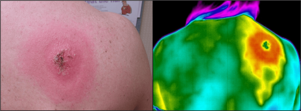
In a case series, Johns Hopkins researchers used infrared thermography (IRT) to capture heat patterns associated with the erythema migrans (EM) rash and surrounding inflammation in early Lyme disease.

The imaging revealed inflammation following venous channel-like patterns that extended well beyond what could be seen with the naked eye, offering new clues about how a Lyme disease infection may spread under the skin.

Images were taken with visible light (A) and infrared (B) cameras in a room-temperature-controlled clinical exam room. The EM and heat extensions (venous channels) are visible by infrared thermography (IRT).

Why was this study done?

Lyme disease, caused by Borrelia burgdorferi bacteria from tick bites, triggers inflammation that increases blood flow, warmth, redness, and swelling in the skin around the bite. When present, the early-stage EM rash can be difficult to identify, especially on darker skin or when the rash is faint, changing, or multiple lesions are present. Missing the rash can delay diagnosis and treatment.

Infrared thermography (IRT) is a non-invasive imaging method that detects skin heat patterns invisible to the human eye. IRT has been used in other inflammatory conditions but not previously tested in Lyme disease. The Johns Hopkins researchers explored whether IRT could reveal visible and hidden aspects of EM, including inflammation patterns that could shed light on how the bacteria and the body’s immune response spread through the skin.

How was this study done?

In this cases series, three patients with suspected early Lyme disease and EM rashes were reported. Researchers used an infrared camera to record skin temperature patterns, comparing rash areas to nearby healthy skin and to visible light photographs. Because inflammation from a Lyme infection increases local blood flow and warmth, thermography could highlight where inflammation extends beyond the visible rash borders. The team also noted whether the thermal patterns aligned with possible venous channels beneath the skin.

What were the major findings?

The study found that the Lyme disease EM rash area showed increased warmth compared to nearby healthy skin, making the rash easier to detect using infrared thermography.

Thermal images revealed heat patterns following vein-like tracks, suggesting inflammation may be spreading along venous channels.

These patterns extended well beyond the visible rash, indicating that underlying inflammation develops beyond visible rash borders.

Such evidence of venous dissemination might help inform hypotheses about how Borrelia burgdorferi spreads through tissue during early infection.

What is the impact of this work?

Although infrared thermography is not available in most clinical practices and is unlikely to change routine Lyme disease diagnosis at this time, it could become a valuable research tool. The ability to visualize venous channel-like and extended dissemination patterns may deepen scientific understanding of how Borrelia burgdorferi infection and inflammation spread in early Lyme disease. It may also help researchers study EM in patients with darker skin tones, where visible rash signs can be harder to detect. Larger studies are needed to confirm these findings and determine IRT’s accuracy, reliability, and potential role in diagnosis across diverse patient groups. If validated, this method could help support earlier and more accurate detection, providing a clearer picture of disease spread and potentially improving early diagnosis, treatment, and patient outcomes.

This research was supported by The Lyme Disease Research Foundation of Maryland

Study team members: Natalia I. Brothers • Alison Rebman • Jonathan M. Zenilman • John Aucott

Receive the Latest News from the Johns Hopkins Lyme Disease Research Center

Receive the Latest News from the Johns Hopkins Lyme Disease Research Center

Join our mailing list to receive the latest news and updates from Johns Hopkins Rheumatology and the Johns Hopkins Lyme Disease Research Center.

Interested In

You have Successfully Subscribed!

Filed Under: Lyme Disease Research, Lyme Disease Research Highlights Tagged With: lyme disease research news

Johns Hopkins Lyme Disease Research Center

Johns Hopkins Lyme Disease Research Center

The Johns Hopkins Lyme Disease Research Center is focused on patient-based research in all manifestations of Lyme disease. Our goal is to translate our pioneering research into improved patient care, education, and health outcomes.

Use of This Site

All information contained within the Johns Hopkins Lyme Disease Research Center website is intended for educational purposes only. Physicians and other health care professionals are encouraged to consult other sources and confirm the information contained within this site. Consumers should never disregard medical advice or delay in seeking it because of something they may have read on this website.

Primary Sidebar

  • About the Center
  • Lyme Disease Information
  • Meet Our Team
  • Our Research
  • Support Our Work

Footer

Johns Hopkins Rheumatology

  • Johns Hopkins Rheumatology
  • Arthritis Center
  • Lupus Center
  • Myositis Center
  • Scleroderma Center
  • Sjögren’s Syndrome Center
  • Vasculitis Center

Connect with Us

  • Facebook
  • Twitter
  • YouTube

U.S. News and World Report Rankings Badge

Johns Hopkins Medicine Johns Hopkins Medicine logo
Language Assistance Available:
  • Español
  • American Sign Language
  • עִברִי
  • אידיש
  • አማርኛ
  • 繁體中文
  • Français
  • Tagalog
  • Русский
  • Português
  • Italiano
  • Tiếng Việt
  • Ɓàsɔ́ɔ̀-wùɖù-po-nyɔ̀
  • Igbo asusu
  • èdè Yorùbá
  • বাংলা
  • 日本語
  • 한국어
  • Kreyòl Ayisyen
  • العربية
  • Deutsch
  • Polski
  • Ελληνικά
  • ગુજરાતી
  • ภาษาไทย
  • اُردُو
  • فارسی
  • हिंदी
  • Deitsch
  • ខ្មែរ
  • မြန်မာ
Contact & Privacy Information
Price Transparency
Notice of Privacy Practices Privacy Statement
Terms & Conditions of Use Non-Discrimination Notice
Manage Cookie Preferences
Copyright © 2025 Johns Hopkins Rheumatology